HHS researcher working on “Google for pathology”
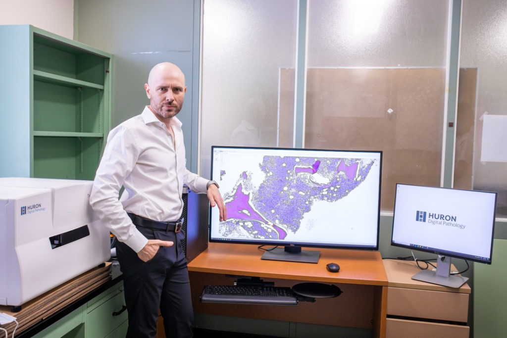
Dr. Clinton Campbell envisions a future where pathologists work as information specialists in sophisticated computer labs with the world’s most current, relevant data at their fingertips. Instead of diagnosing disease one slide at a time under a microscope, there would be thousands of images to compare it to online.
Campbell is working to make that vision possible by harnessing a type of artificial intelligence (AI) known as machine learning.
From microscope to machine
The first steps toward this high-tech future are taking place at Hamilton Health Sciences (HHS) right now thanks to an innovative pilot project exploring digital pathology and AI. It’s the only project of its kind in Canada, and could help modernize the way pathology is conducted around the world.
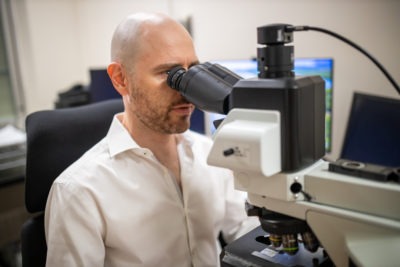
Pathologists currently use a microscope to view sections of human tissue
“Pathologists currently use an old-fashioned light microscope to view sections of human tissue, which is 300-year-old technology,” said Campbell, one of about 100 pathologists in Canada who specializes exclusively in blood disorders like leukemia. Campbell is leading an HHS study into digital pathology and AI. He is supported by Drs. Catherine Ross and Monalisa Sur for this pilot.
Pathologists study tissue samples of disease taken from patients during surgery, or in the case of blood, during a procedure known as a bone marrow biopsy.
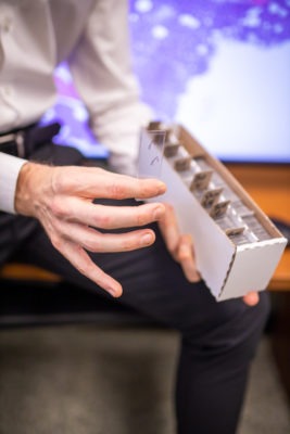
Samples are placed on glass slides
Samples are placed on glass slides for examination under a microscope. Pathologists then look at minute features of cells and tissue and extract information to better understand the disease. This, in turn, may lead to a diagnosis — or at least narrow down to one of a few possible diagnoses and therefore the best treatment options for patients.
Rethinking diagnostic medicine with AI
“The system we have currently works well,” said Campbell. “But we can’t share images of human tissues in real time with colleagues or do `virtual peer review’ where multiple pathologists can examine slides together at the same time.”
Currently, when a pathologist at another hospital wants Campbell to view their slides and offer insights, the slides are shipped to him in thick cardboard folders.
“It would allow for wider, faster consultation.”
“This is an inefficient process that leads to delays in diagnosis. Imagine how much better it would be if we could share these slides digitally in real time,” said Campbell. “It would allow for wider, faster consultation. And if you add AI into the mix, you’ve also got access to a vast amount of knowledge that’s sorted through machine learning to prioritize the very best information to help with your case.”
Machine learning, in the most basic sense, is a type of AI that can automatically learn patterns from data, like pathology images, and then use this knowledge to make useful predictions about the world. This means faster, better reports because machine learning will help extract the most important information, and then compare this to tens, hundreds or even thousands of other previously diagnosed samples instantly.
“It’s kind of like having Google for pathology, with a team of virtual pathologists on standby in the cloud 24/7,” he says. “This could transform the way diagnostic medicine is done.”
The partnership
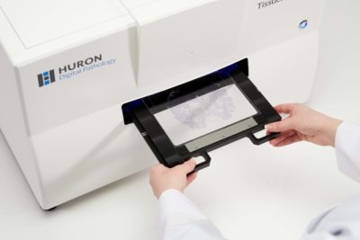
The TissueScope is a scanner that digitizes glass slides at microscopic resolution, allowing researchers and healthcare providers to more easily collaborate, accelerate research and unlock new tools such as those enabled by AI.
HHS is conducting a one-year pilot project on digital pathology in partnership with Huron Digital Pathology in St. Jacobs.
Huron’s AI-based image search platform connects pathologists to the expertise of their colleagues worldwide, increasing the speed and effectiveness of patient care.
HHS is using a scanner provided by Huron to collect and catalogue digital slide images. The project started in May and is based at HHS Juravinski Hospital and Cancer Centre. The goal is to collect 3,000 digital images from blood and bone marrow pathology slides.
The partnership between HHS and Huron was made possible through the Ontario Bioscience Innovation Organization (OBIO), a not-for-profit organization dedicated to advancing health technology innovation and commercialization. The OBIO recently announced the launch of the first eight Early Adopter Health Network (EAHN) innovation projects, including the Huron-HHS partnership.
Finding a digital image solution
So why has it taken so long for pathology to dip its toes into the digital pool? A major hurdle has been image file size and processing.
Hamid Tizhoosh heads the Laboratory for Knowledge Inference in Medical Image Analysis (KIMIA Lab) at Waterloo. He helped Huron develop its technology and has been working with Huron and Campbell to develop a system that will load images faster and more efficiently.
They are doing this by collecting only the small sections of pathology specimen images that relate directly to the disease rather than trying to load the entire image. Images are then processed with AI tools to make a powerful image search engine. It’s not unlike searching using the image of a cat or a dog on Google, where similar images of cats and dogs appear in the results. This is powered by a type of AI. The difference with this technology is that instead of a pet, it involves searching for images of human tissue with a similar disease or diagnosis.
“Huron and the KIMIA Lab piloted this groundbreaking technology, and we’re now expanding this into hematology with the hope of improving it and applying it to blood disorders like leukemia, lymphoma and myeloma,” said Campbell.
Faster, better care for patients
“Groundwork is being laid at HHS that will change the way pathologists do their jobs.”
“This pilot project is an example of how HHS is collaborating with the best and brightest to modernize health care in Canada and around the world,” said Dr. Ted Scott, Vice President, Research & Chief Innovation Officer for HHS. “Groundwork is being laid at HHS that will change the way pathologists do their jobs, and these innovations will pave the way for faster, better care for patients.”
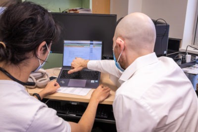
A McMaster University e-health intern student and study Principal Investigator Dr. Clinton Campbell review data used by machine learning software to annotate digital images of bone marrow tissue samples.
Campbell hopes to see a working digital pathology system in place in Hamilton within five years, including overlaying the system with AI. He says the pathology profession will need to embrace this new way of working. There’s currently a shortage of pathologists in Canada, but this new way of practising could help attract a new, tech-savvy generation of “superstars” to the profession.
Digital pathology will also allow these specialists to continue working, should Canada ever be hit by another worldwide event like a pandemic.
“It is clearly the way forward for the field of pathology.”
“I think we escaped the worst of the pandemic here in Canada, fortunately,” said Campbell. “But if another such event struck, pathologists could work remotely. It is imperative we prepare for this now by looking into the future.”
While digital pathology is still several years away, Campbell said it’s definitely coming.
“Pathologists who learn to understand and use digital tools and AI will replace those who don’t,” he says. “It is clearly the way forward for the field of pathology, which is increasingly becoming a field of information specialists. And at the end of the day, it will lead to better patient care because of faster, more reliable and robust diagnoses.”