
Imaging technology for the heart guides procedure to save HHS patient’s leg
Earlier this month, physicians at Hamilton Health Sciences saved a man’s leg by using a procedure normally reserved for looking at blockages in the heart. Collaboration between vascular surgeons and interventional cardiologists at the hospital made it possible — a first in Canada with implications for preventing limb amputations.
“This study could help patients worldwide avoid amputations.”
The procedure was a success, and thanks to the innovative partnership between these physicians, the specialized technique for looking inside a patient’s arteries is now being explored more widely in vascular surgery.
Higher degree of accuracy
Hamilton Health Sciences (HHS) is recognized as a world leader in interventional cardiology – a branch of cardiology that uses catheters to treat heart-related diseases.
Heart patients suspected of having blockages are often sent for an angiogram, where x-ray pictures of coronary arteries and vessels help doctors determine the best treatment options for removing blockages so blood can flow better.
Optical coherence tomography (OCT) takes imaging technology to the next level by using infrared light to capture high-resolution images inside a patient’s arteries, displaying the type and severity of disease in the vessel. These images provide a much higher degree of accuracy during stent implantations to open up vessels, and help physicians determine the very best treatment options so that they can take steps to keep the artery open longer.
“OCT provides far more detailed information than we would get from an angiogram,” says HHS interventional cardiologist Dr. Tej Sheth, adding that OCT technology has been used by HHS’ cardiology department for about 10 years. In fact, HHS is recognized as a top centre internationally for expertise in this technique.
First in Canada
“Until recently, OCT had only been adopted in the heart,” says Sheth.
In partnership with vascular surgeon Dr. Fadi Elias, Sheth used the infrared technology to help guide the recent groundbreaking procedure. Performed on Sept. 4, it was the first time such image-guided surgery had been used in a procedure not involving the heart in Canada.
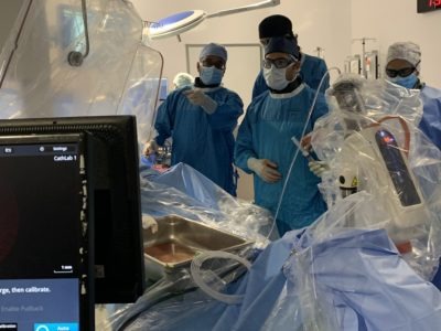
Dr. Tej Sheth (far left) and Dr. Fadi Elias (centre) used optical coherence tomography (OCT) to help guide the recent groundbreaking procedure that saved a man’s leg.
Vascular surgeons treat conditions affecting blood vessels, including those supporting blood flow to the legs and feet.
The procedure took place at HHS’ Hamilton General Hospital in the new hybrid operating suite, which opened last fall and was funded by donors through the Hamilton Health Sciences Foundation. The suite supports leading approaches to cardiac and vascular surgery using integrated imaging technology and surgical equipment.
Treating peripheral artery disease
“I didn’t want to lose my foot or my leg too.”
Retired brick packer Peter Gillie, 69, of Hamilton was the patient for this groundbreaking procedure, which saved his leg from amputation.
“I had lost a toe back in the spring due to poor circulation and I didn’t want to lose my foot or my leg too,” says Gillie. His toe was amputated due to peripheral artery disease, a circulatory problem where narrowed arteries reduce blood flow to the leg and can lead to a diabetic foot, ulcerations, skin breakdown and amputations.
Risk factors include smoking, high blood pressure and high cholesterol. Gillie believes 50 years of smoking likely contributed to his blockage, which also caused leg swelling and a stubborn ulcer. The health scare promoted him to butt out for good five months ago.
“I figured the time had come to quit,” he said.
HHS collaboration
Close collaboration between HHS interventional cardiologists, cardiac surgeons and vascular surgeons started in the trans-catheter aortic valve program, where they work as a team delivering a catheter valve to the heart through blood vessels in the leg. Through discussions, they found some striking similarities in the arteries they treated and the devices, techniques and equipment they used.
OCT technology is optimized for a certain diameter of artery and blood vessel size, which is why it had previously been used exclusively in arteries of the heart. Elias felt that the size of blood vessels in the lower leg were a close enough match to use OCT in vascular surgery. This led to conversations about whether OCT could be used by vascular surgeons when inserting a stent in the blood vessels carrying blood to the legs and feet.
Helping patients worldwide
The collaboration has led to a study using OCT imaging technology for tibial vessel procedures. HHS is one of two centres in North America conducting an evaluation of OCT for the tibial vessels, in partnership with technology manufacturer Abbott Vascular. The other hospital site is in Cleveland, Ohio.
The research project will recruit 50 patients between the two hospital sites. The ultimate goal is to improve limb salvage through improved procedure results and better blood flow, says Elias.
“The culture of collaboration we’ve adopted at HHS is leading the way for innovative, new ways to use the very best technology available to treat our patients,” he says. “This study could help patients worldwide avoid amputations.”