New MRI-ultrasound fusion approach helps find life-threatening prostate cancers
JHCC offers MRI-ultrasound fusion biopsies
New diagnostic imaging technology at Hamilton Health Sciences (HHS) Juravinski Hospital and Cancer Centre is helping to better identify prostate cancer, including life-threatening types, so that patients get the most appropriate treatment.
“When we use this biopsy system, we can target the biopsy to collect samples of cancer that could be life-threatening.” — Dr. Scott Tsai, HHS radiologist
For aggressive prostate cancers, treatment could mean surgery, radiation and/or chemotherapy, while low-risk types may simply need regular monitoring. The JHCC’s new magnetic resonance imaging (MRI) ultrasound biopsy system combines MRI with ultrasound to create 3-D images of the prostate during biopsies.
Prostate cancer
One in eight men will be diagnosed with prostate cancer in their lifetime, making it the most common cancer to affect men in Canada, according to the Canadian Cancer Society.
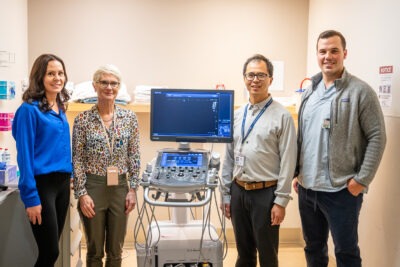
Team members include Jeanine Risk, manager of diagnostic Imaging at JHCC and nuclear medicine; Dr Lori Stewart, staff radiologist and body imaging lead for JHCC; Dr Scott Tsai, radiologist; and Stephen Zuccolo, ultrasound technologist.
Most prostate cancers aren’t considered life-threatening because they grow very slowly and are contained in the prostate gland. For a person diagnosed with a low-risk prostate cancer, the disease may never be an issue over their lifetime, says Dr. Ameya Madhav Kulkarni, an HHS radiologist.
But some types are aggressive and can spread quickly, making them far more dangerous. The best chance for successful treatment is finding prostate cancer early, when it’s still confined to the prostate gland.
Raising the biopsy bar
In Canada, and many parts of the world, a systematic biopsy is the standard of care for helping to diagnose prostate cancer. With a systematic biopsy, a needle is used to take 10 to 12 tissue samples from random parts of the prostate. Systematic biopsies are done using ultrasound to help guide where to take tissue samples from. Ultrasound uses sound waves to produce images, which can provide important information when diagnosing diseases like cancer.
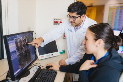
Dr. Ameya Madhav Kulkarni and senior MRI technologist Liz Teixeira
Samples are sent to a lab for testing that includes identifying the type of cancer. But because a systematic biopsy takes samples randomly, there’s a risk of missing cancer.
MRI uses a powerful magnet and radiofrequency waves to make cross-sectional images that can identify aggressive types of prostate cancer, show cancer missed by other tests and help identify the size and location of cancer and whether it has spread.
The new approach of combining MRI and ultrasound creates matched 3-D images of the prostate, allowing radiologists to target areas in the prostate that should be biopsied, thereby increasing the detection of clinically significant cancers.
“The advantage of MRI is that it tends to detect prostate cancers that are clinically significant,” says Dr. Scott Tsai, an HHS radiologist. “When we use this biopsy system, we can target the biopsy to collect samples of cancer that could be life-threatening.”
HHS introduced the technology last November, which included training team members on the new system. HHS and St. Joseph Healthcare Hamilton are the only two hospitals in the region offering MRI-ultrasound fusion biopsies.