
New software a big step forward for pediatric epilepsy unit
Neurologists in the Pediatric Epilepsy Clinic at McMaster Children’s Hospital, Dr. Kevin Jones and Dr. Rajesh RamachandranNair, met with the Hamilton Health Sciences Foundation (HHSF) to advocate for a special software program which allows them to provide better care for patients.
The electroencephalogram (EEG) Source Imaging software, called Curry, can display abnormal brainwave activity as a map on a patient’s MRI scan, allowing the doctor to pinpoint where seizures are happening. This is particularly important with brain surgery, which requires extreme accuracy.
Assessments to pinpoint seizure locations
The Pediatric Epilepsy Clinic is a specialized clinic that provides services for children and teens with epilepsy. As part of their work, doctors assess patients to see if they qualify for surgery to help reduce seizures and the impact on the child’s brain.
During these assessments, doctors Jones and RamachandranNair would inspect the EEG brain waves to find the region where seizures are located to see if the patient qualified for surgery. “It was difficult to pinpoint the exact location where the seizures were coming from,” says Dr. Jones. “We don’t want to remove more brain than we need to.”
It took roughly a year to implement, from pitching the need, to securing donations, to procuring and installing the software. In January, the software became available for the clinic to use.
How it works
The software can display abnormal brainwave activity as a map on a patient’s MRI scan allowing the doctor to pinpoint where seizures are happening. This is particularly important with brain surgery, which requires extreme accuracy.
The software uses complex computer data to analyze brain waves and maps them onto an MRI to help pinpoint where the spikes are.
“We can show the images to families to better explain, and can present the findings to our team of doctors, nurse practitioners, social workers and EEG technologists to discuss each child’s case and determine who is a potential candidate for epilepsy surgery,” says Dr. Jones.
Then, the team meets with the patient and their family to share results and recommendations.
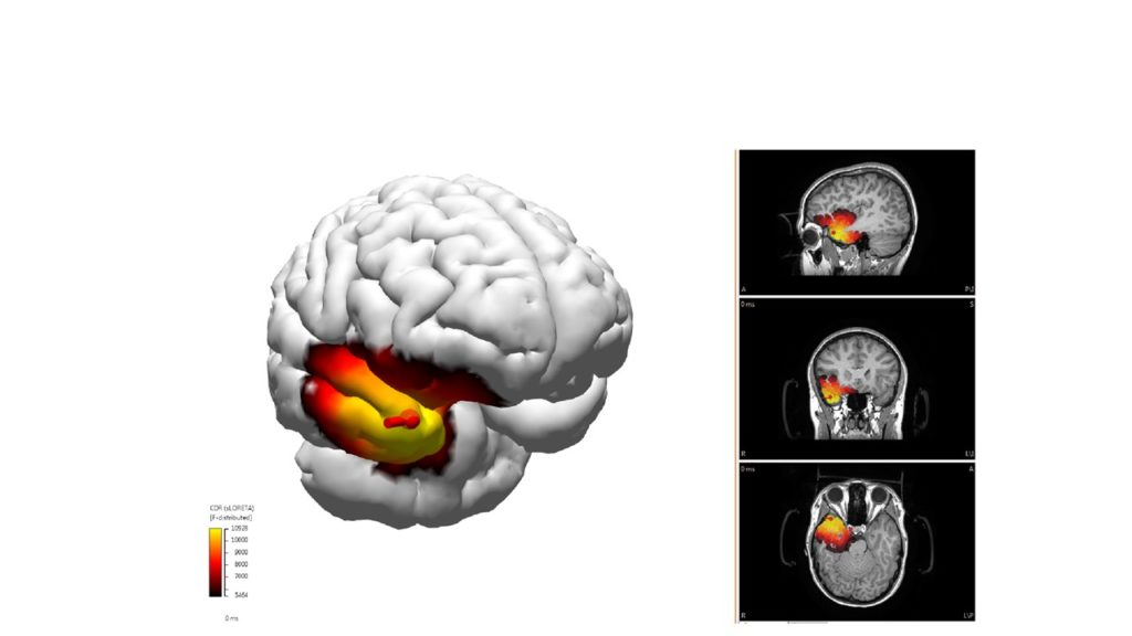
Curry – the EEG Source Imaging software – shows where the patient’s seizures are located.
Curry is now the standard of care for our program. “Not only can we now look at where the spikes come from, but also explore the extent of the seizure network,” Dr. Jones explains.
“There is interest in this worldwide.”
Related:
Up Close: Epilepsy Program